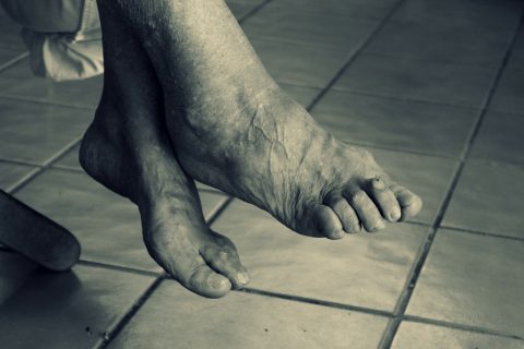
As our feet grow older, they naturally develop more problems. But painful and uncomfortable feet are not something you should have to put up with. A lot can be done to improve comfort, relieve pain and keep you on your feet for life. Below are listed some specific foot health problems that affect an ageing population:
Skin changes As the skin ages, it loses some of its former qualities of elasticity, moisture balance and fatty padding. The skin becomes vulnerable to tears and, therefore, ongoing slower wound healing and infection. The foot is an area particularly vulnerable to skin break-down complications; being at the most distal part of a limb it has susceptibility to peripheral neurological and circulatory loss. A Podiatrist is often the first health professional to thoroughly examine the foot and can be the first to detect skin changes, such as skin cancers, which are more prevalent in the aged foot.
Pressure areas With the average person aiming for 10,000 steps per day, an 80-year-old foot could have tread over 290 million steps in a lifetime. It should therefore come as no surprise to learn that the fatty padding in the foot, either under the heel or the ball of the foot, can be considerably reduced in the ageing patient. The combination of pressure and reduced protection produces pressure-related problems unique to the foot; callouses and corns over bony prominences and metatarsal heads, heel pain from standing and walking, inter-digital neuromas and bursas or capsulitis.
Nail changes Difficulties with bending down, eyesight or focal length and hand grip strength often are the initiating factors for a person to directly contact a Podiatrist for assistance with foot care. Podiatrists regularly treat nails in the aged population and offer professional care of nail pathology such as ingrown nails, fungal nail infections, and wounds related to excessively long or thickened nails.
Changing capability As well as physical changes, there are often cognitive impairments related to chronic disease and complex medical presentations in the aged. Impairment in memory, loss of concentration, impairment in focus and judgment can affect personal care. These mental capacity deficits produce a higher risk profile for the aged foot, which often requires professional input of a Podiatrist as a regular provider of foot care.
Orthopaedic changes The foot shape and appearance can change with ageing due to changes in bony structure and weakness or loss of elasticity in the connective tissues, such as ligaments and tendons. Muscle strains and tendon pathology are common consequences of an active older person who is demanding a lot from an ageing body. Bunions and clawing toes are common presentations in the ageing foot. Other underlying chronic diseases such as arthritis and diabetes often exacerbate foot orthopaedic problems.
Gait changes Falls in the elderly are a concern to people who have experienced falls, their families and the health system at a community level. It has been shown that people at higher risk of falls have a more variable pattern of minimum foot clearance, which could lead to trips and falls. Podiatrists have a role in footwear advice and maintaining the foot to be as pain-free and functional as possible.
Foot pain Foot pain affects up to 24% of people over 65 years of age (4). Pain is associated with altered activities of daily living, balance and gait. Some of the risk factors for pain are gender (with women reporting more foot pain), obesity and chronic health problems.
Podiatrists form an integral part of the health care team for ageing Australians. Podiatrists play a key role in assisting ageing Australians with general foot care, which would otherwise be left unattended and could lead to more serious problems, including infection, hospitalisation and, in worst-case scenario, even amputation.
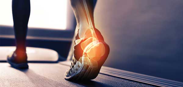
Severs disease is foot pain and/or ankle pain as a result of inflammation of the growth plate of the heel bone in children. In the initial stages of the condition most children displaying signs of Severs disease will tend to hobble or limp off the football field, soccer pitch, basketball court or netball court. Kids will complain of sore heels near the end of activity. This condition most commonly affects children between the ages of 8 to 14 years. This type of condition commonly occurs in those kids who are very active with sport.
The cause of the pain in Severs disease is thought to be the tractional forces applied to the growth plate of the heel bone, Achilles tendon and the plantar fascia. This tractional force by the Achilles tendon and the plantar fascia on the growth plate is often aggravated by tight calf muscles and excessively pronated feet (i.e. feet that “roll in” too far). The good news is that this heel pain in children is very simple to treat and children usually respond very quickly to treatment once treatment of Severs disease commences.
Treatment
When this condition affects both feet, often the diagnosis can be made clinically. If only one foot is affected then x-rays should always be taken of both feet if your child fails to respond to what is considered normal treatment for Severs disease. This is to ensure serious problems such as bone infection or bone tumours are not overlooked. Even in cases where both feet have been affected, x-rays or MRI scans should be carried out if a child is failing to respond to conservative treatment.
Treatment of Severs disease usually involves a combination of ice therapy, activity review and / or modification, review of training surfaces, exercises, footwear review and orthotic inserts where foot function is causing excessive traction on the heel growth plate.
Treatment of Severs disease does NOT require surgery. This foot condition responds very well to conservative treatment within a matter of weeks. If your child suffers from heel pain, get them checked out especially when only one foot is affected.
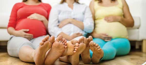
Pregnancy triggers many different changes in a woman’s body, and can lead to problems that affect your feet and legs. Foot pain is one of these complaints that can often be overlooked. Due to the natural weight gain during pregnancy, a woman’s centre of gravity is completely altered. This causes a new weight-bearing stance and added pressure to the knees and feet. Furthermore, the naturally released hormones that prepare the body for child birth also cause relaxation of the ligaments in the feet.
Flat Feet
Hormones increase during pregnancy. Some of these hormones help relax ligaments and other structures to allow a vaginal birth. These same hormones can also relax the ligaments in your feet, leading to flat feet (fallen arches) and over-pronation. This loosening of ligaments can also increase your shoe size during pregnancy. Therefore, you may have to wear a half or whole size larger after you give birth. Your growing womb, baby and breasts contribute to weight gain that causes extra stress on your already compromised feet, especially your arches. It is not uncommon for pregnant women to develop heel pain (plantar fasciitis) because of the extra weight and stress on the arches.
Prevention/Treatment:
Try to avoid standing for long periods of time and walking barefoot. Take a break when you can, and sit down and elevate your feet.
Supportive, properly fitted shoes and arch supports will help; see a Podiatrist to discuss custom orthotics.
Swelling
Oedema (swelling) is an increase in fluid in the tissues of your body. Swelling in your feet and ankles during pregnancy is very common. It is usually caused by an increase in blood volume that occurs to help you carry extra oxygen and nutrients to your baby. Pregnancy hormones can also cause changes in the blood vessels, which may lead to swelling. You may notice that your shoes become too tight. An increase in foot size due to swelling are common and temporary.
Prevention/Treatment:
Do not stand still for long periods of time. Walking gets your calf muscles working, which helps pump some of the extra fluid out of your legs and feet.
Rest several times a day, elevating your feet as much as possible when sitting down.
Wear compression stockings to help decrease the swelling.
Drink plenty of water throughout the day. Try to avoid foods that contain large amounts of salt, as they will increase your fluid retention.
Rest on your left side. This decreases the pressure on blood vessels and allows more fluid to move from your legs to your upper body.
Wear the correct shoe size for your feet.
Toenail Changes
Your toenails tend to grow faster during pregnancy. This is usually due to increased blood volume and circulation of hormones. Because you are providing nutrients for your baby, the cells in your toenails can sometimes be deprived of an adequate amount of nutrients. This can cause the development of nail brittleness, ridges or grooves that go across your nail, and dark or discoloured lines in the nail bed. A nail might even become loose and fall off. These nail changes will usually go away after your pregnancy.
Prevention/Treatment:
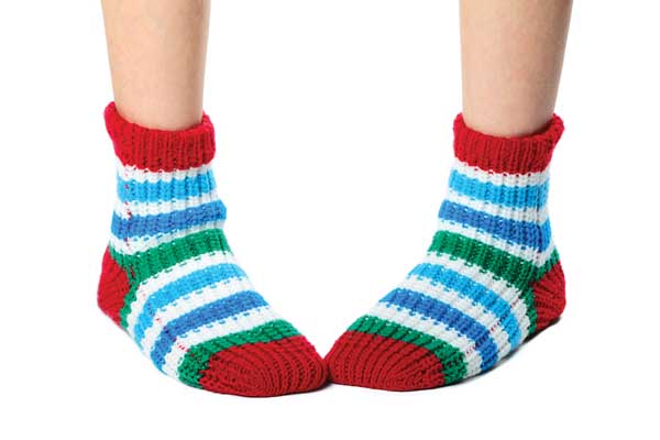
Do not wear shoes or socks that are too tight. The extra pressure they put on the skin around the nails may cause ingrown toenails.
Eat healthy, well-balanced meals. This will help supply the nutrients needed for you and your baby.
Do not trim toenails too short. Swollen skin can overlap the corners of short toenails, causing ingrown toenails.
Have someone else trim your toenails or get a pedicure if you are not able to see/reach your feet.
Podiatrists are able to provide a complete assessment of your feet during pregnancy and provide advice and treatment, should it be required.
Pigeon Toe (Metatarsus Adductus) is a common deformity of the foot generally noticed at birth, though subtle deformity can be overlooked and not picked up till later. It presents with the front part of the foot positioned inward in comparison with the rear of the foot, giving the foot a ‘kidney-shaped’ appearance. The rearfoot and ankle are usually in a normal position. The inside border of the foot appears concave with an exaggerated arch and there is often a wider space between the first and second toes. Metatarsus adductus that is undiagnosed at birth can become more apparent later in life as an in-toed gait.
What causes Metatarsus Adductus?
A single cause remains unclear but genetic and environmental factors have been suggested. These factors include:
Muscle imbalance and soft tissue contractions
Family history of metatarsus adductus
Position of baby in uterus
Sleeping position of baby
Stunted development of foot in utero (8-9wks)
Abnormal muscle insertions in the foot
Has been linked with developmental dysplasia of the hip (DDH)
The incidence is approximately one in 1000 births. It affects boys and girls equally and there is some evidence that first born children are at a greater risk.
Treatment Options
Chosen treatment options for metatarsus adductus will depend on several factors including the age of the child, severity, rigidity, compliance and expectation levels. Metatarsus adductus responds best to early conservative treatment and may resolve spontaneously without treatment in some children.
It is particularly crucial that treatment begins prior to the child commencing walking (ie before 12 months of age).
Treatment options may include:
Manipulation and stretching
Padded shoes
Changing sleeping position of child
Serial casting
Splints and braces
Surgical correction
Or a combination of the above treatments
The most well acknowledged treatment prior to 12 months of age is the use of plaster casts, changed periodically over a 1-3 month period to correct the deformity. Usually if treatment is left until after weight-bearing commences the likelihood of success drops considerably.
Osgood-Schlatter disease causes pain in the front of the knee. It is commonly seen in boys and girls between the ages of 9-16. It features a painful lump just below the knee, this is because of inflammation of the patellar ligament at the tibial tuberosity. The pain usually occurs during physical activity such as running, jumping, squatting and going up and down stairs.
Around 75% of cases affect boys and occurs in up to 20% of sporty children compared to 4% of a group of all activity levels. In a quarter of cases, both knees are affected and it is more likely to occur around periods of rapid growth. As the condition is due to irritation and damage of the growth plate, it can only occur while the growth plate is present, up to the age of 16 years approximately.
Mechanical factors play a big role in Osgood–Schlatter’s disease. When the feet are in ‘perfect’ alignment, the quadriceps muscles, patella tendon, patella and tibial tuberosity are all in a line. Any force created by using the thigh muscles transmits to the tuberosity in a direct, front-on direction. A pronated foot will increase the quadriceps angle in a similar way that a knock kneed position would. The change in the angle of pull can leave the trochanter more vulnerable from an angled pulling force.
Ignoring symptoms or adopting a ‘no pain, no gain’ attitude is likely to cause further damage and prolong recovery in patients with Osgood Schlatters disease. Immediate, appropriate treatment in patients with this condition is essential to ensure a speedy recovery.
Some of the factors which may contribute to the development of Osgood Schlatters disease include:
A sudden increase in training or sporting activity
Inappropriate training
Recent growth spurts
Inappropriate footwear
Muscle tightness or weakness (particularly the quadriceps)
Joint stiffness
Poor lower limb biomechanics
Poor foot posture
Treatment techniques the Podiatrist may use include:
RICE (Rest, Ice, Compression, and Elevation)
Orthotics-The Podiatrist may discuss the use of foot orthotics as part of a treatment plan. This is especially important if there are biomechanical factors that exacerbate the tension on the patella tendon. This will help support the foot and reduce the effect of biomechanical abnormalities such as an excessively pronated foot.
Stretching—Advice on stretching the quadriceps & hamstrings.
Recommence sport in a month or so
Training changes—depending on your activities the podiatrist may recommend modifications to a training regime. Return to activity should be supervised by a qualified person such as a podiatrist or experienced adolescent coach.
The success rate of treatment is largely dictated by patient compliance.
Muscles of your leg, ankle & foot make up about half of the weight of this part of the body and they are required to make even the smallest of movements such as moving your ankle when you walk or run or just tapping your toe. If too much stretch is put through one of your muscles, you may end up with a painful muscle strain.
What causes a muscle strain?
A muscle strain, or a muscle pull occurs when a muscle in your lower limb is overstretched or overworked. Even if the injury from overstretching or overworking occurs more to the attaching tendon it can also be classified under the term muscle strain.
How are muscle strains classified?
There are 3 grades. All muscle strains include tearing of some muscle fibres:
Grade I (mild): Very few muscle fibres have been injured. Pain may not be felt until the following day after the instigating activity. No swelling or bruising is noted.
Grade II (moderate): With this category many muscle fibres are torn which results in a decrease in strength and often limited movement. Some muscle fibres remain uninjured and intact. Pain is present both when stretching the muscle and on muscle strength testing. Swelling and bruising may be noted.
Grade III (severe): All fibres of the muscle are completely torn. Severe swelling, pain, and bruising accompany a grade III strain.
How are muscle strains treated?
The initial approach to Podiatry of your muscle strain will depend on how long after your injury that you seek treatment. The immediate line of defence straight after a muscle strain should be the application of ice and compression, followed by rest and elevation for the affected muscle.
Once the initial pain and inflammation has calmed down, your Podiatrist will focus on improving the flexibility and strength of the involved muscle. Static stretches to increase the flexibility of the muscle will be prescribed by your Podiatrist early on in your treatment as these types of stretches encourage the healing tissues to withstand stretch and they ensure that you do not lose any normal movement.
Rest is also an important part of your Podiatry treatment. ‘Relative rest’ is a term used to describe a scale of resting compared to the normal activity you would be doing.
Along with stretching exercises, your Podiatrist will also prescribe strengthening exercises in order to get your strained muscle back in top shape.
In addition to stretching and strengthening the muscle, taping or wrapping the affected muscle with an elastic bandage may be done by your Podiatrist in order to assist initial swelling, and to provide support to the muscle as you rehabilitate it. As well your Podiatrist may use some of the following approaches to aid in the recovery of your strained muscle: massage, dry needling, joint mobilisation, heat (later) & even custom orthotics in your shoes to aid with correct lower limb alignment which may further aid in preventing re-injury.